|
Biodiversity Data Journal :
General research article
|
|
Corresponding author:
Academic editor: Pavel Stoev
Received: 14 Jul 2015 | Accepted: 13 Aug 2015 | Published: 17 Aug 2015
© 2015 Jackson Means, Elizabeth Francis, Avery Lane, Paul Marek
This is an open access article distributed under the terms of the Creative Commons Attribution License (CC BY 4.0), which permits unrestricted use, distribution, and reproduction in any medium, provided the original author and source are credited.
Citation:
Means J, Francis E, Lane A, Marek P (2015) A general methodology for collecting and preserving xystodesmid and other large millipedes for biodiversity research. Biodiversity Data Journal 3: e5665. https://doi.org/10.3897/BDJ.3.e5665
|
|
Abstract
Background
With an estimated 80% of species remaining undescribed (but see
New information
Here we summarize a methodology for large-bodied millipede collection, curation, and preservation for genetic analyses with the hope that sharing these techniques will stimulate interest in these charismatic detritivores.
Keywords
millipede, detritus, mimicry, evolution, collection, α-taxonomy, DNA, RNA, specimen
Introduction
Millipedes perform an invaluable ecological service in the form of detritus fragmentation and nutrient recycling (
Several methods have been previously outlined for the collection of millipedes (
Material and methods
The following methods have been developed for collecting xystodesmid millipedes in the Appalachian Mountains. They have also been successfully used throughout California and Costa Rica and may serve as a basis for collections-based taxonomic research in other litter-dwelling arthropod groups.
Site Identification
With the exception of desert-dwelling species, millipedes generally lack a waxy epicuticle, rendering them permeable to water and susceptible to desiccation (
Collecting Kit
Our standard field collecting kit (Fig.
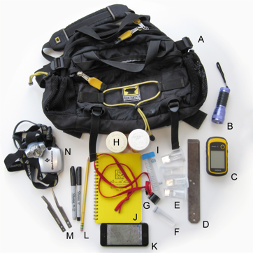
Field collection kit, including a (A) Mountainsmith lumbar pack (8 L volume); (B) UV flashlight; (C) GPS unit; (D) breaker bar; (E) 20 mL collection vials (cell counter type); (F) 100% alcohol-filled vials for millipedes that prematurely die (Sarstedt 8 mL plastic screw-top vials); (G) 10X and 20X Coddington loupes; (H) 100 mL collection vials (large pharmacy pill vial type); (I) 50 mL Falcon tube; (J) Field notebook; (K) Apple iPhone with internal GPS and Gaia GPS app with predownloaded USGS topo quads; (L) pencil, fine and extra fine point permanent Sharpie markers; (M) narrow and wide tip featherweight forceps from Bioquip; (N) headlamp from Black Diamond (Ion model).
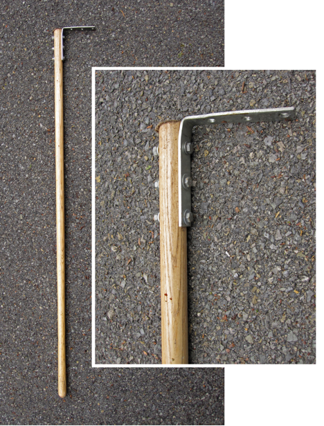
Millipede Rake
Xystodesmid millipedes can often be found beneath leaf litter and as such a tool for the removal of litter is helpful in their collection. The millipede rake, originally adopted by Rowland Shelley (pers. comm.), consists of a wooden broom handle with a metal corner brace bolted to one end (Fig.
Other Useful Equipment
(i) UV flashlight (Fig.
(ii) Gaia GPS app (Fig.
(iii) Breaker bar and headlamp (Fig.
(iv) Coddington loupes (Fig.
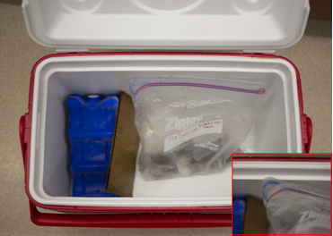
Millipede Storage
Once collected, xystodesmids should be stored in 20 mL plastic cell-counter vials with a piece of moistened moss to reduce desiccation (Fig.
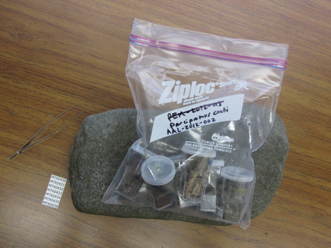
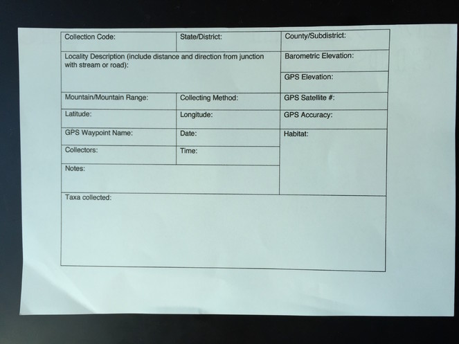
Collection Cards
Supporting geographical information is essential for collections based biodiversity research and a specimen without a locality is nearly useless as a museum specimen. The location data recorded on collection cards (Fig.
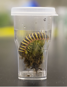
Rearing Immatures & Housing Live Specimens
Occasionally juvenile xystodesmid millipedes, which are soft and not yet fully sclerotized, must be collected due to possession of unique gonopods (indicating a new species or form) or an otherwise interesting biological feature. Juvenile specimens should be kept in the laboratory to develop into an adult. Developing xystodesmids should be stored in glass terrariums or 5 L battery jars with soil, moss and detritus from the habitat where the specimen was collected. As a result, millipedes that are not yet mature may complete their development for preparation and preservation later. Care should be taken not to disturb the specimens (e.g., sifting through the leaves or persistently checking) because individuals that are molting are extremely delicate and the molting chambers in which millipedes undergo ecdysis are fragile and easily damaged.
Live Photography
Millipedes representing new taxa, with unique color patterns, or lacking a photograph, should be documented while alive using a high resolution (≥ 10 megapixels) digital camera (e.g., a Canon EOS 6D with a 65 mm macro lens for smaller genera ≤ 2 cm and a 50 mm macro lens for larger-bodied genera ≥ 2 cm). A flash with a diffuser should be used to produce soft light that will not generate glare (e.g., a Canon Macro Twin Lite MT-24EX flash). Photographs should be captured from above (dorsal habitus) and from the side (lateral habitus) to record color patterns of the tergites, legs, and other external features. In case of millipedes with a color spot on the prozonite, an image of the millipede in a defensive coil should be captured. A bed of moss should be used to reduce light reflection and provide a natural background. Each photograph is assigned a unique code derived from the individual specimen code (e.g., if the specimen code is MPE0003 then the images should be named MPE0003_1, MPE0003_2…). These unique codes link image files with specimens, and are used to track and retrieve images in digital image archives.
DNA & RNA Storage
After returning from the field with live specimens, material should be kept at room temperature (20°C), or in an environmental chamber maintained at 12°C and a light-to-dark setting of 12:12. The following protocol describes methods for the stabilization and storage of tissues for subsequent use in genetic analyses. Before beginning specimen processing, sterilize the bench area with a 10% bleach solution and gather required materials. Prepare one 1.5 mL microcentrifuge tube for each millipede by adding 500 µL of RNAlater (Qiagen, Valencia, California) to each and placing in a tube rack. Use an alcohol resistant pen to record the specimen code on Tough Spots label stickers (USA Scientific, Ocala, Florida) for each of the tubes. Dumont 3C tweezers should be used to dissect legs for storage in RNAlater. To sterilize between processing each specimen, dip the end of the tweezers into 100% ethanol, ignite with a flame, and hold upright until the flame is extinguished. Then, hold the tips of the tweezers in the lower blue region of the flame for 3 s, followed by a rinse of deionized water. Carefully place the sterilized tweezers on a fresh Kimwipe or weigh paper to maintain sterility before use. Fill specimen vials ¾ full (~ 10 mL) with 70% isopropanol. Take precaution to not let any fecal material, soil, parasitic mites, or other organic material contaminate the tissue collected for analysis. If needed, use a moistened Kimwipe (Kimberly-Clark, Roswell, GA) to wipe away debris from arthropods and gloves. Assign a unique specimen code label for each animal (already printed on Resistall paper) and record the code in pencil on the collection record. Grasping the xystodesmid ventral side up between the index and middle fingers and thumb, use the 3C tweezers to dissect legs at their base from the left posterior side of the millipede and remove legs anteriorly to segment 8 (the segment immediately posterior to the gonopods in males, see Fig.
Museum Specimen Curation/ Alcohol Storage
Specimens prepared for long-term museum storage should be kept in 70% isopropanol for two weeks at -20°C in 3 (11.1 mL) or 4 (14.8 mL) dram shell vials (depending on the size of the individual) to allow the alcohol to diffuse into the specimen and reduce brittleness. An initial storage at -20°C for two weeks avoids putrefaction that sometimes results when millipedes are fixed in room-temperature alcohol. After two weeks, alcohol should be replaced and subsequently the material should be stored at room temperature. Depending on the size of the specimen, a second round of isopropanol change may be needed—particularly for large specimens or if the alcohol is darkened to a point where the label is discolored. For museum storage, up to 12 individual shell vials can be stored together in 70% isopropanol kept at room temperature in 473 mL straight-sided glass jars with polypropylene caps and a PTFE cap-liner. The stock caps from individual shell vials should be replaced with a cotton plug (tight enough that the specimen does not fall out if the vial is inverted). This process will ensure diffusion of the isopropanol throughout the jar. Place the vials cotton end down into the jar. Finally, the jar should be topped off with 70% isopropanol. In contrast, field-preserved specimens in ethanol (those which prematurely died in the field) should receive fresh, cold 100% ethanol and remain at -20°C indefinitely.
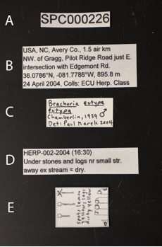
Labels prepared on white chemical resistant paper (e.g., Resistall, Forbon, 100% rag content, Tyvek) should be included with each specimen. Each specimen should have five associated labels. (1) Specimen labels should indicate the unique specimen code assigned to each millipede during processing (Fig.
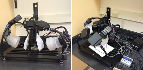
Gonopod Dissection and Examination
For identification and imaging, the left gonopod should be removed using a thin needle clamped in a pin vise (00 pins or minutens work well). Using the pin, the membrane surrounding the left gonopod should be perforated, and the gonopod completely separated from the membrane. After which, the gonopod should be lifted out using size 55 Inox forceps. Special care must be taken when piercing the membrane between the two gonopods; the membrane is notably tougher in this area and if not properly perforated the right gonopod may accidentally be lifted out with the left. To remove, grasp the inter-gonopodal membrane or the coxal apodeme, taking care to avoid damaging the gonopod. The actual gonopod should never be grasped itself to remove from the segment. Dissected gonopods should then be examined to identify the species since they are often the species-characteristic features used for taxonomy. The left gonopod, optionally, can be photographed using a microphotography system (e.g., Canon dSLR with a 65mm macro lens mounted on a Passport II Portable Digital Imaging System, Visionary Digital, Charlottesville, VA, Fig.
Discussion
The collection and preservation methods presented here are an effective and efficient means of collecting millipedes of the family Xystodesmidae and other terrestrial arthropods, such as centipedes, beetles, and spiders. These methods represent a demonstrably successful model and starting point for investigators interested in collection-based research with most ground-dwelling arthropods, and as such may be modified for other taxa. For example, we have implemented these methods for the collection and preservation of ground beetle specimens and their DNA. Rather than storing specimens in alcohol, however, beetles are pinned and labeled with acid-free 100% cotton archival paper. In addition, these methods can be adapted for individual use and to fit the preexisting workflow of the lab.
Our methodology for hand collecting xystodesmid millipedes was developed to provide a reliable and useful protocol for collection based biodiversity research. Recent studies of millipedes using these techniques and subsequent molecular phylogenetics have shed light on the evolutionary relationships of the Diplopoda, but much remains to be explored solely in terms of an α-taxonomic and primary descriptive standpoint (
Acknowledgements
We are grateful for funding provided by a Virginia Tech USDA NIFA Hatch Project (VA-160028) and a National Science Foundation Phylogenetic Systematics award (DEB#1410911). We thank Tim McCoy for his assistance in the field and with lab start-up. Katy Lawler and Nina Zegler provided useful assistance both in the field and laboratory. We thank Bill Shear and two anonymous reviewers for suggestions that greatly improved the manuscript.
References
- Harnessing the world's biodiversity data: promise and peril in ecological niche modeling of species distributions.Annals of the New York Academy of Sciences1260(1):66‑80. https://doi.org/10.1111/j.1749-6632.2011.06440.x
- Millipede Taxonomy after 250 Years: Classification and Taxonomic Practices in a Mega-Diverse yet Understudied Arthropod Group.PLoS ONE7(5):e37240. https://doi.org/10.1371/journal.pone.0037240
- Global Biodiversity: Indicators of Recent Declines.Science328(5982):1164‑1168. https://doi.org/10.1126/science.1187512
- Darwin’s legacy to rove beetles (Coleoptera, Staphylinidae): A new genus and a new species, including materials collected on the Beagle’s voyage.ZooKeys379:29‑41. https://doi.org/10.3897/zookeys.379.6624
- The water relations and cuticle of Paradesmus gracilis (Diplopoda, Strongylosomidae).Quarterly Journal of Microscopical Science91:453‑464.
- Diversity in Tropical Rain Forests and Coral Reefs.Science199(4335):1302‑1310. https://doi.org/10.1126/science.199.4335.1302
- Natural History Collections as Emerging Resources for Innovative Education.BioScience64(8):725‑734. https://doi.org/10.1093/biosci/biu096
- Desert millipedes: a rationale for their distribution. In Myriapod biology.Academic Press,London,171-181pp.
- Millipedes as model detritivores.Berichte des Naturwissenschaftlich-Medizinischen Verein Innsbruck10:277‑288.
- 21 years of shelf life between discovery and description of new species.Current Biology22(22):R943‑R944. https://doi.org/10.1016/j.cub.2012.10.029
- Diplopoda. In Arachnida and Myriapoda.Pensoft,Sofia-Moscow,505-533pp.
- Effect of calcareous road dust on land snails (Gastropoda: Pulmonata) and millipedes (Diplopoda) in acid forest soils of the Daniel Boone National Forest of Kentucky, USA.Forest Ecology and Management186:177‑183. https://doi.org/10.1016/s0378-1127(03)00259-7
- Lewis J, Pursell F (1997) The Nature Conservancy at work in the Indiana karst: The bioinventory of the subterranean fauna of the Blue River Bioreserve. In: Stitt R (Ed.) : 1997 Karst and Cave Management Symposium 13th National Cave Management Symposium.
- A revision of the Appalachian millipede genus Brachoria Chamberlin, 1939 (Polydesmida: Xystodesmidae: Apheloriini).Zoological Journal of the Linnean Society159(4):817‑889. https://doi.org/10.1111/j.1096-3642.2010.00633.x
- Phylogenetic systematics of the colorful, cyanide-producing millipedes of Appalachia (Polydesmida, Xystodesmidae, Apheloriini) using a total evidence Bayesian approach.Molecular Phylogenetics and Evolution41(3):704‑729. https://doi.org/10.1016/j.ympev.2006.05.043
- A Mullerian mimicry ring in Appalachian millipedes.Proceedings of the National Academy of Sciences106(24):9755‑9760. https://doi.org/10.1073/pnas.0810408106
- A species catalog of the millipede family Xystodesmidae (Diplopoda: Polydesmida), Virginia Museum of Natural History.Special Publications17(1):1‑117.
- Bioluminescent aposematism in millipedes.Current Biology21(18):R680‑R681. https://doi.org/10.1016/j.cub.2011.08.012
- Relative efficiency of pitfall trapping and hand-collecting from plots for sampling of millipedes.Biodiversity and Conservation4(4):429‑439. https://doi.org/10.1007/bf00058426
- The biodiversity data knowledge gap: Assessing information loss in the management of Biosphere Reserves.Biological Conservation173:74‑79. https://doi.org/10.1016/j.biocon.2013.11.020
- Estimating subterranean species richness using intensive sampling and rarefaction curves in a high density cave region in West Virginia.Journal of Cave and Karst Studies66:39‑45.
- The geological record and phylogeny of the Myriapoda.Arthropod Structure & Development39:174‑190. https://doi.org/10.1016/j.asd.2009.11.002
- Current Status of the Myriapod Class Diplopoda (Millipedes): Taxonomic Diversity and Phylogeny.Annual Review of Entomology52(1):401‑420. https://doi.org/10.1146/annurev.ento.52.111805.090210
- The influence of millipedes on selected soil elements: a microcosm study on three species occurring on coastal sand dunes.Functional Ecology15(1):51‑59. https://doi.org/10.1046/j.1365-2435.2001.00493.x
- Development of an optimal sampling protocol for millipedes (Diplopoda).Journal of Insect Conservation10(3):277‑288. https://doi.org/10.1007/s10841-006-6699-z
- The symbiotic mites of some Appalachian Xystodesmidae (Diplopoda:Polydesmida) and the complete mitochondrial genome sequence of the mite Stylochyrus rarior (Berlese) (Acari:Mesostigmata:Ologamasidae).Invertebrate Systematics23(5):445. https://doi.org/10.1071/is09036
- Sternites and spiracles – The unclear homology of ventral sclerites in the basal millipede order Glomeridesmida (Myriapoda, Diplopoda).Arthropod Structure & Development43(1):87‑95. https://doi.org/10.1016/j.asd.2013.11.003
Supplementary material
An editable Microsoft Word file of the collection card.