|
Biodiversity Data Journal :
Taxonomic paper
|
|
Corresponding author:
Academic editor: Jason Bond
Received: 27 Dec 2014 | Accepted: 06 Sep 2015 | Published: 07 Sep 2015
© 2015 Recep Özkütük, Mert Elverici, Yuri Marusik, Kadir Kunt
This is an open access article distributed under the terms of the Creative Commons Attribution License (CC BY 4.0), which permits unrestricted use, distribution, and reproduction in any medium, provided the original author and source are credited.
Citation:
Özkütük R, Elverici M, Marusik Y, Kunt K (2015) A new species of Harpactea Bristowe, 1939 from Turkey (Araneae: Dysderidae). Biodiversity Data Journal 3: e4419. https://doi.org/10.3897/BDJ.3.e4419
|
|
Abstract
A new species of Harpactea Bristowe, 1939, H. alanyana sp. n. is described from southern Turkey. The new species appears closely related to H. osellai Brignoli, 1978. Detailed description and illustrations of the new and related species are provided. The relationships of the two species are discussed.
Keywords
Alanya, Antalya, Mediterranean, spider, woodlouse hunters
Introduction
Harpactea Bristowe, 1939 is large genus of dysderid spiders that includes 172 species distributed in the Mediterranean region from the Iberian Peninsula to Turkmenistan (
The goal of this article is to describe a recently discovered species of Harpactea from Turkey on the basis of both sexes.
Materials and methods
Specimens were collected from Antalya Province in the Mediterranean region of Turkey, using a sifter. The specimens were preserved in 70% ethanol and deposited in the Anadolu University Zoology Museum. Digital images of the copulatory organs were taken with a Leica DFC295 digital camera attached to a Leica S8AP0 stereomicroscope and 5-15 photographs were taken in different focal planes and combined using automontage software. SEM microphotographs were made from dried and sputter coated (by gold) organs by use of a Zeiss Ultra Plus SEM device (Anadolu University, Eskişehir). All measurements are in mm, with methods as per (
Abbreviations
The following abbreviations are used in the text: Carapace and abdomen: AL, abdominal length; CL, carapace length; CWmax, maximum carapace width; CWmin, minimum carapace width. Eyes: AME, anterior median eyes; PLE, posterior lateral eyes; PME, posterior median eyes; AMEd, diameter of anterior median eyes; PLEd, diameter of posterior lateral eyes; PMEd, diameter of posterior median eyes. Chelicera: ChF, length of cheliceral fang; ChG, length of cheliceral groove; ChL, total length of chelicera (lateral external view). Legs: Ta, tarsus; Me, metatarsus, Ti, tibia; Pa, patella; Fe, femur; Tr, trochanter; C, coxa; D, dorsal; Pl, prolateral; Rl, retrolateral; V, ventral.
Depository
AUZM, Anadolu University Zoology Museum, Eskişehir, Turkey; MCSNV, Museo Civico di Storia Natuale di Verona, Italy; NHMG, The Natural History Museum of Geneva, Switzerland; AZM, Alaşehir Zoological Museum, Manisa, Turkey; ZMMU, Zoological Museum, Moscow Lomonosov State University, Russia.
Taxon treatment
Harpactea alanyana, sp. n.
-
scientificName: Harpactea alanyana; class:Arachnida; order:Araneae; family:Dysderidae; nomenclaturalCode:ICZN; genus:Harpactea; specificEpithet:alanyana; continent:Asia; country:Turkey; countryCode:TR; stateProvince:Mediterranean; county:Antalya; municipality:Alanya; locality:Taşatan Plateau; verbatimLatitude:36°38'37.3500"; verbatimLongitude:032°04'42.0900"; verbatimCoordinateSystem:degrees minutes seconds; samplingProtocol:sifter; eventDate:24 April 2011; habitat:pine forest; sex:1 male; lifeStage:adult; preparations:whole animal (ETOH); recordedBy:R.S. Özkütük; disposition:in collection; institutionCode:AUZM; basisOfRecord:PreservedSpecimen
-
scientificName: Harpactea alanyana; class:Arachnida; order:Araneae; family:Dysderidae; nomenclaturalCode:ICZN; genus:Harpactea; specificEpithet:alanyana; continent:Asia; country:Turkey; countryCode:TR; stateProvince:Mediterranean; county:Antalya; municipality:Alanya; locality:Asmaca Village; verbatimLatitude:36°36'32.3000"; verbatimLongitude:032°03'12.4000"; verbatimCoordinateSystem:degrees minutes seconds; samplingProtocol:sifter; eventDate:3 January 2013; habitat:pine forest; sex:1 male, 1 female; lifeStage:adult; preparations:whole animal (ETOH); recordedBy:M. Elverici; disposition:in collection; institutionCode:NHMG; basisOfRecord:PreservedSpecimen
-
scientificName: Harpactea alanyana; class:Arachnida; order:Araneae; family:Dysderidae; nomenclaturalCode:ICZN; genus:Harpactea; specificEpithet:alanyana; continent:Asia; country:Turkey; countryCode:TR; stateProvince:Mediterranean; county:Antalya; municipality:Alanya; locality:Asmaca Village; verbatimLatitude:36°36'32.3000"; verbatimLongitude:032°03'12.4000"; verbatimCoordinateSystem:degrees minutes seconds; samplingProtocol:sifter; eventDate:3 January 2013; habitat:pine forest; sex:2 female; lifeStage:adult; preparations:whole animal (ETOH); recordedBy:M. Elverici; disposition:in collection; institutionCode:AUZM; basisOfRecord:PreservedSpecimen
-
scientificName: Harpactea alanyana; class:Arachnida; order:Araneae; family:Dysderidae; nomenclaturalCode:ICZN; genus:Harpactea; specificEpithet:alanyana; continent:Asia; country:Turkey; countryCode:TR; stateProvince:Mediterranean; county:Antalya; municipality:Alanya; locality:Avsallar Town; verbatimLatitude:36°38'21.5000"; verbatimLongitude:031°45'24.9000"; verbatimCoordinateSystem:degrees minutes seconds; samplingProtocol:sifter; eventDate:6 January 2013; habitat:pine forest; sex:1 male, 1 female; lifeStage:adult; preparations:whole animal (ETOH); recordedBy:K.B. Kunt; disposition:in collection; institutionCode:ZMMU; basisOfRecord:PreservedSpecimen
-
scientificName: Harpactea osellai Brignoli, 1978; namePublishedIn:Brignoli P.M. 1978. Ragni di Turchia V. Specie nuove o interessanti, cavernicole ed epigee, di varie famiglie (Araneae). Revue suisse de Zoologie. Vol.85. P.461-541.; taxonomicStatus:accepted; class:Arachnida; order:Araneae; family:Dysderidae; genus:Harpactea; specificEpithet:osellai; country:Turkey; countryCode:TR; stateProvince:Amasya; locality:Borabay Lake; eventDate:4 June 1969; institutionCode:MCSNV
Description
Measurements [Holotype ♂ / Paratype ♀]: AL 1.88 / 3.00; CL 1.70 / 2.28; CWmax 1.30 / 1.72; CWmin 0.63 / 0.92; AMEd 0.08 / 0.11; PLEd 0.07 / 0.09; PMEd 0.05 / 0.08; ChF 0.28 / 0.40; ChG 0.25 / 0.27; ChL 0.64 / 0.93. Leg measurements are given in (Table
|
Leg |
Fe |
Pa |
Ti |
Me |
Ta |
Total |
|
I |
1.48 / 1.88 |
0.83 / 1.14 |
1.18 / 1.55 |
1.00 / 1.35 |
0.40 / 0.45 |
4.89 / 6.37 |
|
II |
1.35 / 1.75 |
0.75 / 1.13 |
1.13 / 1.45 |
1.10 / 1.38 |
0.38 / 0.38 |
4.71 / 6.09 |
|
III |
1.07 / 1.45 |
0.57 / 0.73 |
0.83 / 0.78 |
1.10 / 1.38 |
0.34 / 0.48 |
3.91 / 4.82 |
|
IV |
1.56 / 2.00 |
0.75 / 0.98 |
1.38 / 1.53 |
1.41 / 1.78 |
0.50 / 0.53 |
5.60 / 6.82 |
No apparent dimorphism between the sexes except in body sizes. Carapace hexagonal, reddish brown, dull and smooth. Carapace covered with very short, tiny and sparsely distributed setae. AME, PLE and PME closely grouped; AME separated (Fig.
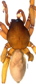
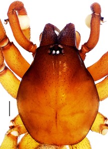
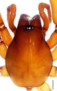
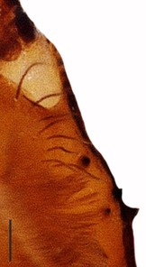
Cheliceral groove with four teeth; at retromargin, with a small tooth located at the base of the groove, and with a more developed second tooth a little above the second quarter. Both retromarginal teeth conical and tubercular. Promarginal teeth more strongly developed; the one closer to the base of the cheliceral groove larger and almost twice the size of the other (Fig.
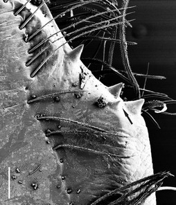
Legs yellowish brown, covered with tiny brownish hairs on all surface. Leg formula IV, I, II, III. Tarsi with three claws. Paired claws toothed. Paired claws of leg I and II with 7 teeth; leg III and IV with 4 teeth. Scopula weak and in ventral position on the first distal half on 3rd and 4th tarsi and on 4th metatarsus; relatively strongly developed on 3rd metatarsus in ventral position at the first distal half. Coxae III with 1 prolateral spine; coxae IV with 1-2 prolateral spines. Patellae III with 1 spine dorsally. Further details on leg spination are given in (Table
|
♂ |
Leg I |
Leg II |
Leg III |
Leg IV |
|
C |
0 |
0 |
1 Pl |
1 Pl |
|
Fe |
2 Pl |
1, 1 Pl |
1, 1 D 1, 1, 1 Rl |
1, 2 D |
|
Pa |
0 |
0 |
1 D |
0 |
|
Ti |
0 |
0 |
1, 1 Pl 1, 1, 1 Rl 1, 1, 2 V |
1, 1, 1 Pl 1, 1, 1 Rl 1, 1, 2 V |
|
Me |
0 |
0 |
1, 1 Pl 1, 1, 1 Rl 2, 1, 2 V |
1, 1, 1 Pl 1, 1, 1, 1 Rl 1, 1, 2 V |
|
♀ |
||||
|
C |
0 |
0 |
1 Pl |
1-2 Pl |
|
Fe |
2 Pl |
1, 1 Pl |
1, 1 D 1, 1 Rl |
1, 1 Pl 1, 1 D |
|
Pa |
0 |
0 |
1 D |
0 |
|
Ti |
0 |
0 |
1, 1 Pl 1, 1, 1 Rl 1, 1, 2 V |
1, 1, 1 Pl 1, 1, 1 Rl 1, 1, 2 V |
|
Me |
0 |
0 |
1, 1 Pl 1, 1, 1 Rl 2, 1, 2 V |
1, 1, 1 Pl 1, 1, 1, 1 Rl 1, 1, 2 V |
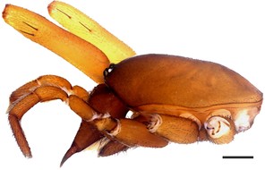
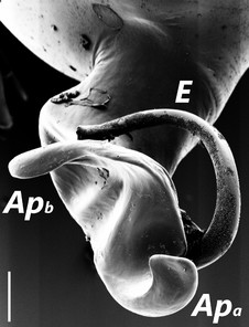
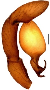
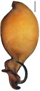
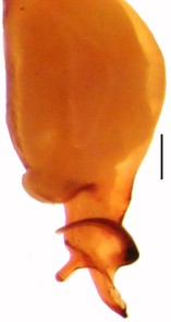
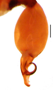
Bulbus almost oval, yellowish brown, with embolus and two apophyses. An apparent membranous part between the bulb and the distal appendages; embolus hook-shaped, black, almost homogenously sclerotized. Bent anteriorly following the same course as apophysisb. Apophysisa L-shaped, apically well sclerotized, short and strong. Apophysisb separated from the membranous part of the bulbus with a wide angle from the base and orientates anteriorly. Apically blunt, conical and in the shape of a triangular apophysis in retrolateral view (Fig.
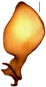
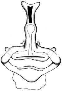
Distal expansion of spermatheca tenuously developed between distal crest and rod-shaped part of the anterior spermatheca. Nevertheless, it is more sclerotized at the surface compared to other parts of the anterior spermatheca. Distal crest gradually tapers through the tip. Rod-shaped part of the anterior spermatheca almost one and a half times the length of the distal crest. Basal transverse part of the anterior spermatheca widens through laterals and narrows through the tips, in the shape of open wings. A ring-shaped strongly sclerotized structure is apparent at the juncture of the rod-shaped part of the anterior spermatheca and the basal transverse part of the anterior spermatheca. Transverse bar in the shape of a lip. Posterior diverticulum prominent, and in the form of a broad membranous sac (Fig.
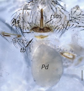
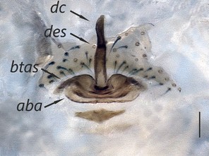
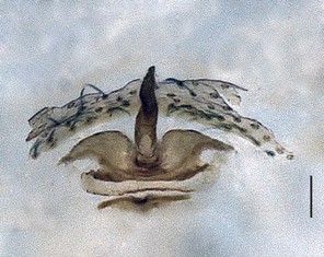
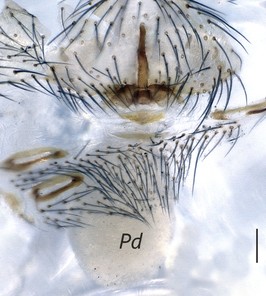
Harpactea alanyana sp. n. Abbreviations: As, anterior spermatheca; aba, anterior basal arc; btas, basal transverse part of the anterior spermatheca; dc, distal crest; des, distal expansion of the spermatheca; Pd, posterior diverticulum; rsas, rod-shaped part of the anterior spermatheca; tb, transverse bar. Scale lines: 0.02 mm
b: Ditto
c: Ditto
d: Ditto, ventral view
Diagnosis
Harpactea alanyana sp. n. can be easily distinguished from all known species of Harpactea by the unique structures of the male and female copulatory organs. The male palp of the new species is similar to that of H. osellai. However, the two species can be easily separated by the shape of the bulb; by having a less curled embolus compared to H. osellai and by the shape of the distal apophysis (Fig.
Etymology
The specific name refers to the type locality (Alanya District, Antalya Province, Turkey).
Distribution
Harpactea alanyana sp. n. is currently known only from the type locality and its vicinity.
Ecology
All specimens were collected from slopes and ridges lying parallel to the coastline, from sea level up to a maximum of around 1200 m on peaks of the Taurides Mountain range, by sifting tree litter of mixed forest with the following species Pinus nigra, Quercus coccifera, Arbutus andrachne, Ceratonia siliqua, etc. Adult males are known to be active from late autumn until the middle of spring, while females are only known from the winter.
Taxon discussion
H. alanyana;
1. Based on the oval bulbus, the massive embolus and apophysisa,
2. the presence of spines on the patellae and coxae,
"H. alanyana" belongs to "D. group rubicunda" by the characters stated above.
Notes
Acknowledgements
We are deeply indebted to Dr. Fulvio Gasparo (Trieste, Italy) who discussed differences between males of the new species and H. osellai in those days when the first male specimen was collected, and then gave good advice and encouraged us to describe the new species together with the female; to Dr. Peter Schwendinger (Geneva, Switzerland) and Francesco Ballarin (Beijing, China) who shared their knowledge on the collections where the type specimens of Harpactea osellai are kept; and also to Dr. Rahşen Kaya (Bursa, Turkey) who photographed the holotype specimen of Harpactea osellai for our use. Dr. Müjdat Çağlar (Eskişehir, Turkey) provided assistance during SEM photography and Mr. Savaş Kunt aided during field studies (Antalya, Turkey). The English of the text was kindly checked by Dr. David Penney (Manchester, United Kingdom).
References
- Le Harpactea (Araneae, Dysderidae) della fauna italiana e considerazione sullaloro origine.Atti della Accademia Gioenia di Scienze naturali in Catania18:190‑221. [InItalian].
- Il genere Dasumia Thorell (Araneae, Dysderidae), sua nuova definizione e revisione delle specie italiane.Memorie del Museo Civico di Storia Naturale di Verona14:465‑486. [InItalian].
- The Checklist of the Spiders of Turkey.2014. URL: http://www.spidersofturkey.info
- Ragni di Turchia IV. Leptonetidae, Dysderidae ed Agelenidae nuovi o interessanti di grotte della Turchia meridionale (Araneae).Quaderni di Speleologia, Circolo Speleologico Romano3:37‑54. [InItalian].
- Ragni di Turchia V. Specie nuove o interessanti, cavernicole ed epigee, di varie famiglie (Araneae).Revue suisse de Zoologie85:461‑541. [InItalian]. https://doi.org/10.5962/bhl.part.82243
- Spiders from Turkey, VI. Four new species from the coast of the Black Sea (Araneae).Bulletin of the British Arachnological Society4:310‑313.
- Taxonomic revision of the epigean representatives of the spider subfamily Harpacteinae (Araneae: Dysderidae) on the island of Crete.Zootaxa1169:1‑32.
- The genus Rhode and the harpacteine genera Stalagtia, Folkia, Minotauria, and Kaemis (Araneae, Dysderidae) of Yugoslavia nad Crete, with remark on the genus Harpactea.Revue Arachnologique10(6):105‑135.
- Araneiden, Opilionen und Chernetiden. In Penther, A. und E. Zederbauer, Ergebnisse einer naturwissenschaftlichen Reise zum Erdschias-Dagh (Kleinasien).Annalen des Naturhistorischen Museums in Wien20:114‑154.
- World Spider Catalog.15.5.Natural History Museum Bern. Release date:2014-12-20. URL: http://wsc.nmbe.ch