|
Biodiversity Data Journal :
Taxonomic Paper
|
|
Corresponding author: Cheng-Bin Wang (entomologist@qq.com)
Academic editor: M. Andrew Johnston
Received: 19 Aug 2021 | Accepted: 30 Nov 2021 | Published: 01 Dec 2021
© 2021 Jiang Zhu, Cheng-Bin Wang, Bao-Ying Feng
This is an open access article distributed under the terms of the Creative Commons Attribution License (CC BY 4.0), which permits unrestricted use, distribution, and reproduction in any medium, provided the original author and source are credited.
Citation:
Zhu J, Wang C-B, Feng B-Y (2021) Taxonomical study on the newly-recorded genus Falsonnannocerus Pic from China (Coleoptera, Tenebrionidae, Stenochiinae). Biodiversity Data Journal 9: e73232. https://doi.org/10.3897/BDJ.9.e73232
|
|
Abstract
Background
The genus Falsonannocerus Pic, 1947 (Coleoptera, Tenebrionidae, Stenochiinae, Cnodalonini) includes 13 known species occurring in West Africa, South Asia and Southeast Asia. The bionomics of species in this genus are unknown.
New information
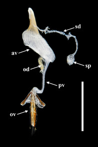
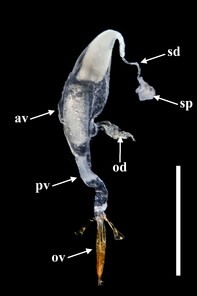
The genus Falsonnannocerus Pic, 1947 is recorded for the first time from China, including two species: F. thailandicus Masumoto, 1986 from Yunnan and F. haizhuensis sp. n. from Guangdong. Morphological characters of the two species are illustrated and compared. The photographs of female reproductive system, which were never shown for this genus before, are also presented.
Keywords
darkling beetle, Cnodalonini, new species, newly-recorded species, taxonomy, Oriental Region
Introduction
The genus Falsonannocerus Pic, 1947 belongs to the tribe Cnodalonini (Coleoptera, Tenebrionidae, Stenochiinae) and was described by
In the present paper, we describe or re-describe two Falsonannocerus species: F. thailandicus Masumoto, 1986 from Yunnan Province, southwest China and F. haizhuensis sp. n. from Guangdong Province, southeast China, representing the first record of the genus from China. Important distinguishing morphological characters of the two species are illustrated with colour plates. The female reproductive system of the genus is provided.
Materials and methods
Specimens were relaxed and softened in hot water for 24 hours, then transferred to distilled water to clean, observe and dissect. In order to examine the genitalia of both sexes, the abdomens were detached and treated with a 10% solution of potassium hydroxide (KOH) for 12 hours, then transferred to distilled water to flush the remaining KOH and stop any further bleaching. After examination, the body parts were mounted on a glass slide with Euparal Mounting Medium for future studies. Photographs of the habitus were taken using microlens on a Canon 5D IV. Detailed photographs with flashlight were performed using a Olympus 10X microlens with a Canon 5D IV. The final deep focus images were created with Zerene Stacker 1.04 stacking software. Adobe Photoshop CS6 was used for post-processing. The terminology adopted in the paper for ovipositor and female genital tubes follows
The material examined for this study is deposited in the following private and public collections: CJZG: collection of Jiang Zhu, Guangzhou, Guangdong, China; CDYZ: collection of Deyao Zhou, Shanghai, China; MYNU: insect collection of Mianyang Normal University, Mianyang, China; NSMT: National Science Museum, Tsukuba, Japan.
Measurement criteria in millimetres (mm) are as follows: antennal length: length between the base of scape and the apex of ultimate antennomere; body length: length between the apices of mandibles and the elytral apices along the mid-line; elytral length: length between the basal border and the apex of elytra along suture; elytral width: widest part of both elytra combined; head length: length between the anterior margin of epistoma and the anterior margin of pronotum along the mid-line; head width: widest part of head (including eyes); pronotal length: length of the pronotum along the mid-line; pronotal width: widest part of pronotum.
Taxon treatments
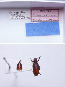
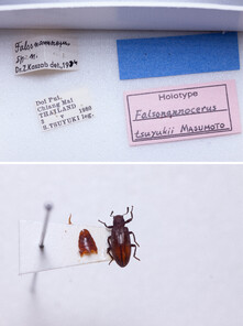
Falsonannocerus thailandicus
-
country: Thailand; verbatimLocality:Chiang Dao; year:1980; month:5; day:2; individualCount:1; sex:male; recordedBy:S. Fukuda; identifiedBy:K. Masumoto; institutionCode:NSMT
-
country: China; stateProvince:Yunnan; verbatimLocality:Yingjiang County, Nongzhang Town [盈江县,弄璋镇]; year:2021; month:4; individualCount:3; sex:2 males, 1 female; recordedBy:Gui-Chang Liu; collectionCode:CDYZ
Description
Male. Body 8.7 mm in length, 3 times as long as wide, widest slightly behind middle of elytra. Lengths of body parts (mm): head (1.2), eye (0.3), antenna (1.8), pronotum (1.7), elytra (5.8); width: head (1.4), eye (0.3), pronotum (1.7), elytra (2.6).
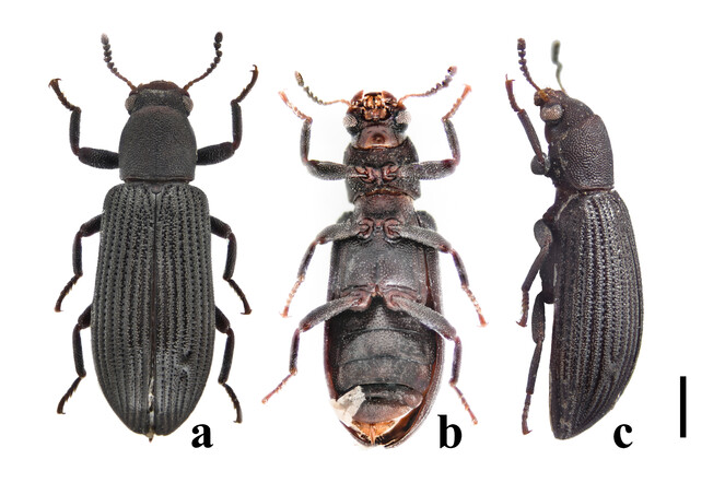
Habitus (Figs
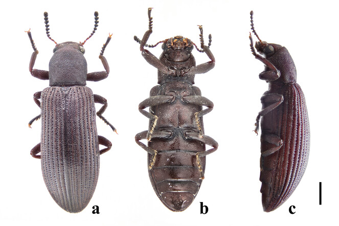
Head transversely elliptical, widest at eye level, strongly convex posteriorly, almost wholly covered with dense and coarse punctures. Epistoma rather small, crescent. Genae oblique, with outer margins rounded. Eyes large, convex laterally, with strong inner ocular sulci. Gula (Fig.
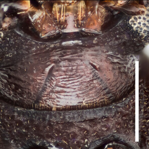
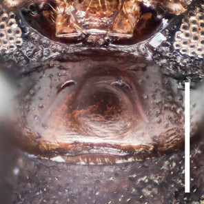
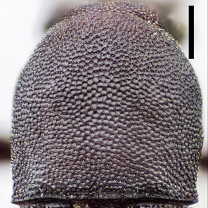
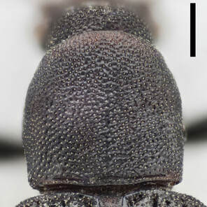
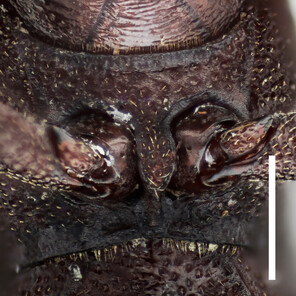
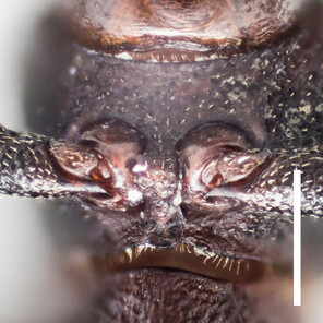
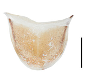
Gulae, pronotums and prosternal processes of Falsonannocerus spp. Scale bar = 0.5 mm.
b: gula of F. haizhuensis sp. n.
c: pronotum of F. thailandicus Masumoto, 1986
d: pronotum of F. haizhuensis sp. n.
e: prosternal process of F. thailandicus Masumoto, 1986
f: prosternal process of F. haizhuensis sp. n.
Mouthparts. Labrum liguliform, surface microreticulate. Maxillary palpi with terminal palpomere securiform. Labial palpi with terminal palpomere elongate, conical. Mentum hippocrepiform, with 6 setae. Submental peduncle trapezoidal.
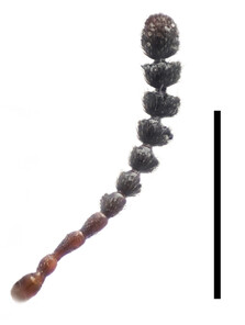
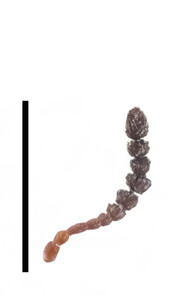
Antennae (Fig.
Prothorax. Pronotum (Fig.
Scutellar shield linguiform, rounded at apex. Disc densely and minutely tuberculate.
Elytra elongate, 2.2 times as long as widest part, widest at apical 3/7. Elytra strongly convex in lateral view, especially in apical half. Lateral margins gradually widened from humeri to apical 3/7, then gradually narrowing to rounded apices. Each elytron with nine irregular rows of close and coarse punctures and short scutellary row in basal 1/6; intervals feebly convex and densely covered with small tubercles throughout. Epipleura wide at base, narrowing towards apex and terminating near apex and sparsely and finely punctate. Hind wings fully developed. Mesoventrite weakly convex towards middle, sparsely punctate, denser posteriorly. Mesepisternum and mesepimeron both triangular, densely punctate. Metaventrite densely punctate and finely grooved along mid-line in posterior half. Metepisternum rather long and thin, densely punctate.
Legs. Femora weakly dilated. Tibiae straight, more or less clavate; protibiae slightly bent near apex of lower side. Tarsi stout. Femora and tibiae densely and coarsely punctate. Setae longer and denser in lower sides of all legs.
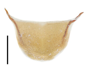
Abdomen. Abdominal sternites III–VI transverse, almost equal in length, densely punctate; intercoxal process on sternite III large, subtriangular; sternite VII semicircular, widely rounded at posterior margin; sternite VIII (Fig.
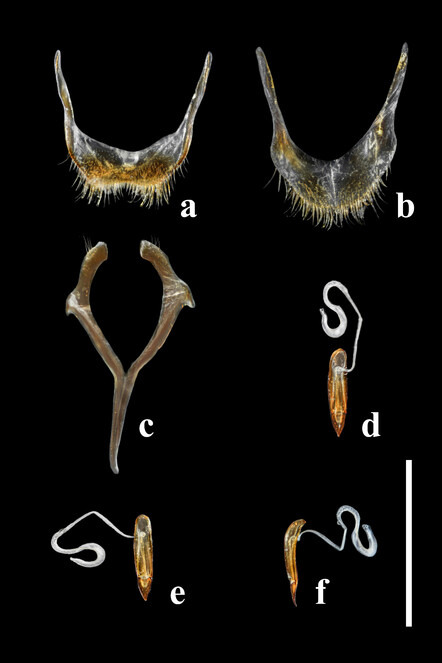
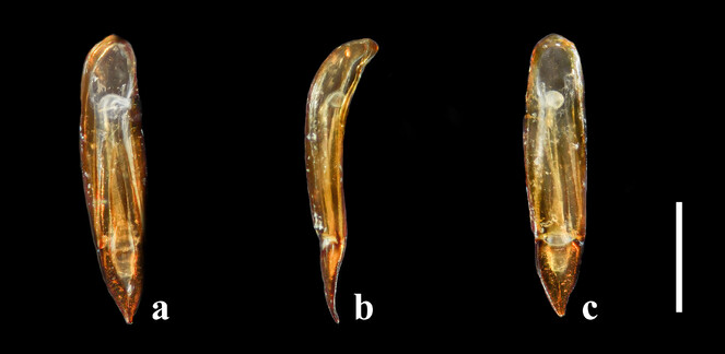
Aedeagus (Fig.
Female. Almost same as male in general appearance. Abdominal tergite VII (Fig.
Distribution
China (Yunnan), Thailand.
Falsonannocerus haizhuensis , sp. n.
-
country: China; stateProvince:Guangdong; verbatimLocality:Guangzhou, Haizhu National Wetland Park [海珠国家湿地公园]; verbatimElevation:8 m; year:2021; month:6; day:9; individualCount:1; sex:female; recordedBy:Jiang Zhu & Wen-Ting Chen; institutionCode:MYNU
Description
Holotype female. Body 6.2 mm in length, 2.5 times as long as wide, widest slightly behind middle of elytra. Lengths of body parts (mm): head (0.9), eye (0.3), antenna (1.2), pronotum (1.2), elytra (4.1); width: head (1.1), eye (0.2), pronotum (1.2), elytra (1.8).
Habitus (Fig.
Head transversely elliptical, widest at eye level, strongly convex posteriorly, almost wholly covered with dense and coarse punctures. Epistoma rather small, crescentic. Genae oblique, with outer margins rounded. Eyes large, convex laterally, with strong inner ocular sulci. Gula (Fig.
Mouthparts. Labrum liguliform, surface microreticulate. Maxillary palpi with terminal palpomere securiform. Labial palpi with terminal palpomere elongated conical. Mentum hippocrepiform, with 3–4 setae. Submental peduncle trapezoidal.
Antennae (Fig.
Prothorax. Pronotum (Fig.
Scutellar shield linguiform, rounded at apex. Disc densely and minutely wrinkled.
Elytra elongate, 2.3 times as long as widest part, widest at apical 3/7. Elytra strongly convex in lateral view, especially in apical half. Lateral margins gradually widened from humeri to apical 3/7, then gradually narrowing to rounded apices. Each elytron with nine irregular rows of close and coarse punctures and short scutellary row in basal 1/6; intervals feebly convex and sparsely covered with small tubercles throughout. Epipleura wide at base, narrowing towards apex and terminating near apex and sparsely and finely punctate. Hind wings fully developed. Mesoventrite weakly convex towards middle, sparsely punctate, denser posteriorly. Mesepisternum and mesepimeron both triangular, densely punctate. Metaventrite densely punctate and finely grooved along mid-line in posterior half. Metepisternum rather long and thin, densely punctate.
Legs. Femora weakly dilated. Tibiae straight, more or less clavate; protibiae slightly bent near apex of lower side. Tarsi stout. Femora and tibiae densely and coarsely punctate. Setae longer and denser in lower sides of all legs.
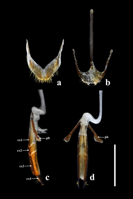
Abdomen. Abdominal sternites III–VI transverse, almost equal in length, densely punctate; intercoxal process on sternite III large, subtriangular; sternite VII semicircular, widely rounded at posterior margin; sternite VIII (Fig.
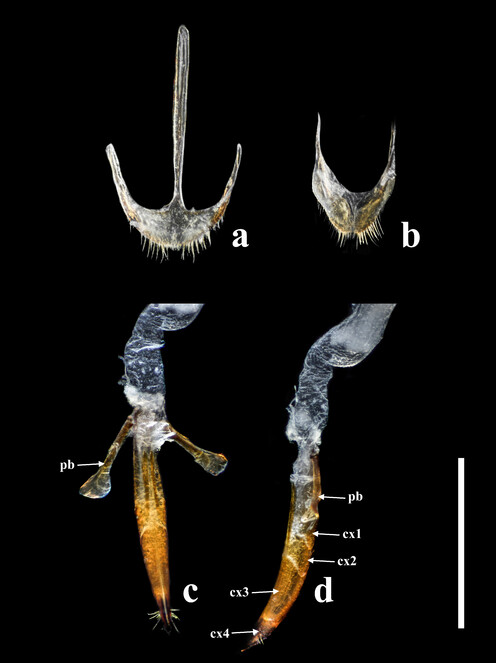
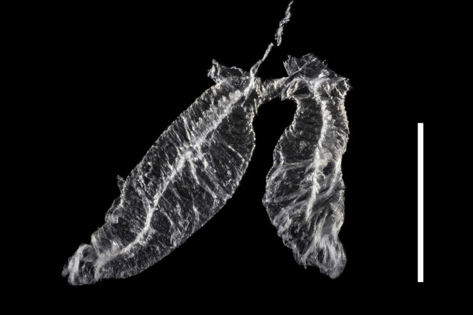
Ovipositor (Fig.
Male. Unknown.
Diagnosis
Except for Falsonannocerus thailandicus Masumoto, 1986 re-described above and F. tsuyukii Masumoto, 1986 from Thailand, F. haizhuensis sp. n. is readily differentiated from other congeners by the combination of the following characters: body colour is without colourful metallic lustre; pronotum is subcylindrical, with its length equal to width; intervals of puncture rows on elytra are covered with small tubercles throughout. For the former two species, the new species can be distinguished from them by the following characters:
In F. thailandicus, body colour is mostly reddish-brown, dull (Figs
In F. tsuyukii, body colour is mostly reddish-brown, lustrous (Fig.
Etymology
The specific epithet is from the Chinese name (Pinyin) of the type locality “Haizhu National Wetland Park”. The name is an adjective.
Distribution
China (Guangdong).
Acknowledgements
We would like to appreciate Shi-Jun Guo (Guangdong, China) and Haizhu National Wetland Park for their support in our work. We acknowledge De-Yao Zhou (Shanghai, China) for his valuable advice, which facilitated our work greatly. Our appreciation is due also to Sui-Jun Lu (Guangdong, China), Hao Xu (Sichuan, China) and Lyu-Bing Zhang (Guangdong, China) for their help to our study. We are obliged to Shûhei Nomura (Japan), Zhi-Zhao Ye (Guangdong, China), Dong-Hao Chen (Guangdong, China) and Yue Wu (Guangdong, China) for helping with speciemen collection. We are grateful to Kojun Kanda (USA), Maxim Nabozhenko (Russia) and Enrico Ruzzier (Italy) who provided constructive comments on an earlier version of the manuscript.
References
- New and little known species of Tenebrionidae (Coleoptera) from Borneo (3).Stuttgarter Beiträge zur Naturkunde A, (N.S.)6:175‑181.
- Neue Tenebrioniden (Coleoptera) Aus Sri Lanka I.Acta Zoologica Academiae Scientiarum Hungaricae26:123‑196.
- Angaben zur Kenntnis der Tenebrioniden Nordvietnams (Coleoptera).Annales Historico-Naturales Musei Nationalis Hungarici72:169‑221.
- Tenebrionidae of East Asia. (I) Tenebrionid beetles from South Sumatra Collected by Mr. Hiroshi Makihara in 1983.Elytra13(1):1‑18.
- Study of Asian Tenebrionidae, I. New species of the Cnodalonine genera of Allopezus, Chaetopsia and Falsonannocerus.The Entomological Review of Japan41(1):1‑26.
- New tenebrionid beetles from East Asia (Coleoptera, Tenebrionidae).Japanese Journal of Systematic Entomology4(2):305‑319.
- Nouveaux coléoptères de la Côte d'Ivoire.Bulletin de la Société Entomologique de France51(10):150‑151. https://doi.org/10.3406/bsef.1946.15925
- Comparative anatomy of the defensive glands, ovipositors and female genital tubes of tenebrionid beetles (Coleoptera).International Journal of Insect Morphology and Embryology9:321‑368. https://doi.org/10.1016/0020-7322(80)90009-4