|
Biodiversity Data Journal :
Taxonomic Paper
|
|
Corresponding author: Carla Rego (crego@fc.ul.pt)
Academic editor: Torsten Dikow
Received: 26 Nov 2021 | Accepted: 28 Jan 2022 | Published: 21 Mar 2022
© 2022 Carla Rego, John Smit, António Aguiar, Délia Cravo, Andreia Penado, Mário Boieiro
This is an open access article distributed under the terms of the Creative Commons Attribution License (CC BY 4.0), which permits unrestricted use, distribution, and reproduction in any medium, provided the original author and source are credited.
Citation:
Rego C, Smit J, Aguiar AF, Cravo D, Penado A, Boieiro M (2022) A pictorial key for identification of the hoverflies (Diptera: Syrphidae) of the Madeira Archipelago. Biodiversity Data Journal 10: e78518. https://doi.org/10.3897/BDJ.10.e78518
|
|
Abstract
Background
Syrphid flies are important ecological indicators and provide crucial ecosystem services, being important pollinators and biological control agents of insect pests. These charismatic insects are conspicuous and, due to their size and colourful patterns, are relatively easy to identify. However, the lack of user-friendly literature (e.g. photographic guides) for most areas may hamper its wider selection as a study group in biodiversity and ecological studies. The syrphid fauna of Madeira Archipelago comprises 26 species, including four endemics (Eumerus hispidus Smit, Aguiar & Wakeham-Dawson, 2004; Melanostoma wollastoni Wakeham-Dawson, Aguiar, Smit, McCullough & Wyatt, 2004; Myathropa usta, Wollaston, 1858 and Xanthandrus babyssa, Walker, 1849), but, despite the current good taxonomic knowledge on this group, information on species distribution, ecology and conservation is still lacking. Here, we provide a pictorial key to the adult hoverflies of Madeira Archipelago highlighting diagnostic characteristics and present photographs of both males and females (in dorsal and lateral views) in colour plates. The key and plates will help researchers to differentiate these species, thus encouraging the use of this insect group in future bioindication studies. In addition, this study also aims to engage a broader audience of non-experts in improving the knowledge on the distribution and ecology of Madeira syrphids.
New information
We provide a checklist for the hoverflies of Madeira Archipelago and a pictorial key to help on species identification.
Keywords
Flower flies, Macaronesia, Madeira endemics, photographic guide, species identification, syrphids, taxonomic key
Introduction
Syrphids, commonly known as hoverflies or flower flies, belong to a large family of flies (Diptera: Syrphidae) with over 6,000 known species (
The syrphid fauna of Madeira has been studied since the mid-nineteenth century by several authors who contributed to a better understanding of species diversity and distribution in this Archipelago (
During the last decades, there has been a growing interest in biodiversity conservation by the general public that has extended to several charismatic invertebrate groups, such as dragonflies and butterflies. In oceanic islands, like Madeira, invertebrate conservation needs to be fostered by engaging researchers, decision-makers and common citizens in knowing, valuing, protecting and making public the unique diversity of life forms of these ecosystems. This interplay is urgent since the biodiversity of oceanic islands worldwide is under threat due to various factors (e.g. land-use change, invasive species, climate change) and, jointly with significant declines in endemic species abundance, many human-driven extinctions have been documented in these unique ecosystems, including in Madeira Archipelago (
Here, we aim to provide a user-friendly pictorial key for the identification of Madeira’s hoverflies, a charismatic bioindicator and ecologically-important insect group. The key was designed for use by non-experts and, altogether with the photos of male and female specimens of all known species occurring in the Archipelago, aims to engage a diverse audience in improving current knowledge on these conspicuous flies.
Materials and methods
Study area
Madeira Archipelago is located in the Atlantic Ocean, nearly 600 km from the African coast (Morocco) and 450 km north from the Canary Islands, between latitudes 32°24′ and 33°07′N and longitudes 16°16′ and 17°16′W. The Archipelago is formed by three groups of volcanic islands and islets: Madeira, Porto Santo and the Desertas Islands (
Laboratory work
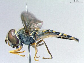
Specimens, both males and females, from all the known species reported to Madeira Archipelago were studied under a stereomicroscope. Most of the specimen’s images were taken with a Leica M125 motorised stereomicroscope, equipped with a IC80 HD digital camera and LAS-Leica Application Suite 3.8 Software. For image stacking, we used the LAS Module “Multifocus” and post-processed the images in ®Adobe Photoshop CC. We also used a Canon 7D digital slr camera with a Canon EF 100 mm 2.8 L Is USM macro lens to capture the habitus of some specimens. The study specimens are deposited in the entomological collections of the Laboratório de Qualidade Agrícola (ICLAM) (Madeira, Portugal) and Naturalis Biodiversity Center, Natural (RMNH) (Leiden, the Netherlands).
Identification keys
|
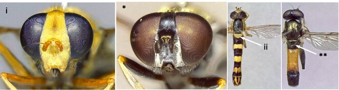
Key to the hoverflies of the Madeira Archipelago The taxonomic key to the adult stages of hoverfly species of the Madeira Archipelago relies on pictorial information to ease interpretation of characters and includes information on morphological differences between males and females. Additionally, photos of male and female specimens of all species (in dorsal and lateral views) are presented in colour plates. In the dichotomous key, the couplet leads present one or more morphological characteristics indicated with symbols (e.g. asterisks for one lead and Roman numerals for the other) which help to easily identify those characteristics in the associated figures. The terminology of morphological characters used in this key follows |
||
| 1 | Face entirely yellow (i); scutellum always yellow, clearly lighter than scutum (ii) (Fig. |
2 |
| – | Face dark or yellow with median dark stripe, sometimes obscured by dense pollinosity (*); scutellum never yellow, sometimes orange-brown (**) (Fig. |
15 |
| 2 | Very large species, over 18 mm, with hornet-like appearance; scutum with yellow markings on anterior half and orange-red colouration on posterior half; metafemur with a small tooth apicoventrally (i); wings with a yellow tinge along the costa (ii) (Fig. |
Milesia crabroniformis |
| – | Smaller species, at most 15 mm, never with a hornet-like appearance; if yellow markings present on scutum, then restricted to lateral margins and scutum never with orange-red markings; metafemur never with a small tooth apicoventrally; wings without yellow tinge along costa | 3 |
| 3 | Thoracic pleura with distinct yellow markings (i); scutum with distinct yellow bands laterally (ii) (Fig. |
4 |
| – | Thoracic pleura without yellow markings (*); scutum without distinct yellow bands laterally (**) (Fig. |
6 |
| 4 | Abdomen distinctly margined (i); male with tooth-like protuberance on metatrochanter (ii) (Fig. |
Ischiodon aegyptius |
| – | Abdomen not margined (*); male without tooth-like protuberance on metatrochanter (Fig. |
5 |
| 5 | Scutum with yellow lateral band restricted to the anterior part of wing base (i); abdomen of males about as long as wings when folded; smaller species: 5-8 mm (Fig. |
Sphaerophoria rueppelli |
| – | Scutum with yellow lateral band uninterrupted, continuing posteriorly of wing base (*); abdomen of males clearly longer than wings when folded (**); larger species: 7-12 mm (Fig. |
Sphaerophoria scripta |
| 6 | Abdomen more or less parallel-sided, as broad as scutum (i) (Fig. |
7 |
| – | Abdomen clearly broadening and oval shaped, clearly broader than scutum (*) (Fig. |
8 |
| 7 | Abdomen with ‘double bands’ on t3, t4 (i) (Fig. |
Episyrphus balteatus |
| – | Abdomen without ‘double bands’, with oblique yellow spots (*), sometimes connected to form bands (Fig. |
Meliscaeva auricollis |
| 8 | Wing vein R4+5 in basal half of cell r4+5 almost parallel to M, curving upwards in apical part (i); eyes pilose (ii); frons distinctly swollen, more obvious in males (iii); larger species: 10-15 mm (Fig. |
9 |
| – | Wing vein R4+5 more or less straight, converging from vein M from the base of cell r4+5 (*); eyes bare, except S. torvus (**); frons not swollen (***); smaller species: 7-13 mm (Fig. |
11 |
| 9 | T3 and t4 with slender yellow or white lunulate maculae, clearly constricted in the middle (i), yellow or white markings covering less than half the length of t3 (Fig. |
10 |
| – | T3 and t4 with larger yellow maculae, which are at most slightly constricted in the middle (+), yellow markings covering more than half the length of t3 (Fig. |
Scaeva albomaculata |
| 10 | Abdominal spots yellow in live specimens; spots on t3 with hind edges curved, their outer and inner corners equally close to anterior edge of the tergite (*) (Fig. |
Scaeva selenitica |
| – | Abdominal spots almost white in live specimens; spots on t3 with hind edges straight and oblique, their outer corners distinctly further removed from anterior edge of tergite than inner corner (ii) (Fig. |
Scaeva pyrastri |
| 11 | Scutum pollinose and dull (i); abdomen with relatively slender yellow bands on t3-t4 (ii); ventral calypter with long erect pili on dorsal surface (iii) (Fig. |
12 |
| – | Scutum pollinose, but clearly shining (*); abdomen typically with yellow spots on t3-t4, sometimes connected to form bands (**); ventral calypter lacking long erect pili on dorsal surface (***) (Fig. |
13 |
| 12 | Eyes pilose, sparse and short in females (i); wing cell BM entirely covered by microtrichia (ii) (Fig. |
Syrphus torvus |
| – | Eyes bare (*); wing cell BM basal ¼ bare (**) (Fig. |
Syrphus vitripennis |
| 13 | Face in frontal view at least as broad as one eye (i); femora at least partially black at the base (ii) (Fig. |
14 |
| – | Face in frontal view clearly narrower than one eye (*); femora entirely yellow (**); abdomen normally with broad yellow maculae, sometimes connected to form bands (Fig. |
Eupeodes nuba |
| 14 | Scutellum predominantly yellow pilose (i); abdominal maculae reaching lateral margins of tergites, normally with spots, but frequently connected to form bands (ii); male with larger genitalia (iii) (Fig. |
Eupeodes corollae |
| – | Scutellum predominantly black pilose (*); abdominal maculae not reaching lateral margins of tergites, usually with spots, only rarely connected to form bands (**); male with smaller genitalia (***) (Fig. |
Eupeodes luniger |
| 15 | Wing vein R4+5 with a strong dip in the cell below (i); larger species (10-16 mm), sometimes with metallic bronze luster, but often with a bee-like appearance (Fig. |
16 |
| – | Wing vein R4+5 without a strong dip in the cell below (*); smaller species (4-12 mm), never with a bee-like appearance (Fig. |
19 |
| 16 | Eyes spotted (i). Entire body largely with metallic bronze luster (Fig. |
Eristalinus aeneus |
| – | Eyes never spotted, but either striped or concolorous. Body without bronze luster | 17 |
| 17 | Eyes striped (*) (Fig. |
Eristalinus taeniops |
| – | Eyes concolorous, without stripes (+) (Fig. |
18 |
| 18 | Eyes with bands of pili (i); wing cell R1 closed (ii) (Fig. |
Eristalis tenax |
| – | Eyes without bands of pili; wing cell R1 open (**) (Fig. |
Myathropa usta |
| 19 | Face in profile with a facial tubercule (i) (Fig. |
20 |
| – | Face in profile more or less straight (*), sometimes mouth-edge clearly protruding (**) (Fig. |
23 |
| 20 | Face entirely black (i); abdomen either entirely dark or with orange-yellow spots; larger species: 5-12 mm (Fig. |
21 |
| – | Face creamy yellow with a black facial stripe (*); abdomen black or partially red; very small species: 4-6 mm (Fig. |
Paragus mundus |
| 21 | Abdomen oval, clearly broader than scutum (i); female with abdomen entirely black or with very small, rounded spots (i); male with broad orange-yellow spots on t3 and t4 (ii), which are sometimes connected (Fig. |
Xanthandrus babyssa |
| – | Abdomen slender and parallel-sided, as broad as scutum (*) (Fig. |
22 |
| 22 | Abdomen entirely black in males (**), at most with reduced orange markings in the female (*); larger species: 7-10 mm (Fig. |
Melanostoma wollastoni |
| – | Abdomen with clear orange markings, triangular on t3 and t4 in females (+), rectangular in males (++); smaller species: 5-8 mm (Fig. |
Melanostoma mellinum |
| 23 | Abdomen clearly petiolate, t2 constricted in basal half (i); smaller species: 5-6 mm (Fig. |
Neoascia podagrica |
| – | Abdomen never that clearly petiolate, t2 never constricted in basal half, abdomen more or less parallel-sided. Larger species: 6-13 mm (Fig. |
24 |
| 24 | Abdomen with a broad orange band (*); larger species: 10-13 mm (Fig. |
Xylota segnis |
| – | Abdomen without orange; smaller species 6-10 mm (+) (Fig. |
25 |
| 25 | Thoracic pleura not dusted (i) and scutum with a pair of longitudinal pollinose vittae, almost reaching scutellum (ii); legs entirely black (iii); abdomen with pollinose spots on t2-t4 (iiii) (Fig. |
Eumerus hispidus |
| – | Thoracic pleura heavily dusted, continuing on the frontal half of the lateral side of scutum (*); legs bicoloured (**); abdomen with yellow spots on the lateral sides of t2 and t3, t4 with dusted areas (Fig. |
Syritta pipiens |


Analysis
The hoverfly species of Madeira Archipelago
The syrphid fauna of Madeira Archipelago comprises 26 species, all considered to be native to these islands (Table
List of the Syrphidae species from Madeira, their distribution in the Archipelago (M – Madeira Island, PS – Porto Santo Island and surrounding islets, D – Desertas Islands) and representative photos of adult males and females (in dorsal and lateral views).
|
Species |
M |
PS |
D |
Photos |
|
Episyrphus balteatus (De Geer, 1776) |
● |
● |
● |
Fig. |
|
Eristalinus aeneus (Scopoli, 1763) |
● |
● |
Fig. |
|
|
Eristalinus taeniops (Wiedemann, 1818) |
● |
Fig. |
||
|
Eristalis tenax (Linnaeus, 1758) |
● |
● |
● |
Fig. |
|
Eumerus hispidus Smit et al., 2004 |
● |
● |
Fig. |
|
|
Eupeodes corollae (Fabricius, 1794) |
● |
● |
● |
Fig. |
|
Eupeodes luniger (Meigen, 1822) |
● |
● |
● |
Fig. |
|
Eupeodes nuba (Wiedemann, 1830) |
● |
Fig. |
||
|
Ischiodon aegyptius (Wiedemann, 1830) |
● |
● |
● |
Fig. |
|
Melanostoma mellinum (Linnaeus, 1758) |
● |
● |
Fig. |
|
|
Melanostoma wollastoni Wakeham-Dawson et al., 2004 |
● |
Fig. |
||
|
Meliscaeva auricollis (Meigen, 1822) |
● |
Fig. |
||
|
Milesia crabroniformis (Fabricius, 1775) |
● |
Fig. |
||
|
Myathropa usta (Wollaston, 1858) |
● |
Fig. |
||
|
Neoascia podagrica (Fabricius, 1775) |
● |
Fig. |
||
|
Paragus mundus Wollaston, 1858 |
● |
● |
Fig. |
|
|
Scaeva albomaculata (Macquart, 1842) |
● |
● |
Fig. |
|
|
Scaeva pyrastri (Linnaeus, 1758) |
● |
● |
● |
Fig. |
|
Scaeva selenitica (Meigen, 1822) |
● |
Fig. |
||
|
Sphaerophoria rueppellii (Wiedemann, 1830) |
● |
● |
Fig. |
|
|
Sphaerophoria scripta (Linnaeus, 1758) |
● |
● |
Fig. |
|
|
Syritta pipiens (Linnaeus, 1758) |
● |
● |
Fig. |
|
|
Syrphus torvus Osten-Sacken, 1875 |
● |
Fig. |
||
|
Syrphus vitripennis Meigen, 1822 |
● |
Fig. |
||
|
Xanthandrus babyssa (Walker, 1849) |
● |
Fig. |
||
|
Xylota segnis (Linnaeus, 1758) |
● |
Fig. |


Acknowledgements
We acknowledge the financial support from Fundação para a Ciência e a Tecnologia (FCT, Portugal) through project PTDC/BIA-BIC/1013/2014, UIDB/00329/2020-2024 and contract DL57/2016/CP1375/CT0001 to MB. We also thank Instituto das Florestas e da Conservação da Natureza (IFCN) for allowing the collection of specimens in Madeira Archipelago.
References
- Eristalinus taeniops (Wiedemann, 1818) (Diptera: Syrphidae) new to Madeira Island, Portugal.Dipterists Digest12:20‑21.
- Islets of Porto Santo: a treasure to be preserved.Serviço do Parque Natural da Madeira,98pp.
- Episyrphus balteatus as a predator of the aphid Sitobion avenae on winter wheat.Entomologia Experimentalis et Applicata42:271‑277. https://doi.org/10.1111/j.1570-7458.1986.tb01032.x
- Britain’s Hoverflies.Princeton University Press, Princeton,312pp. https://doi.org/10.1515/9781400866021
- Zur Kenntnis der Syrphidenfauna des Madeira-Archipels.TenDenZenSupplement:115‑134.
- Dipteren der Insel Madeira.Mitteilungen aus dem Zoologischen Museum in Berlin4:181‑206.
- Neue Dipteren Meiner Sammlung.Mitteilungen aus dem Zoologischen Museum in Berlin10:1‑93.
- Diptères nouveaux ou peu connus. 24e partie. Syrphidi (2e partie).Annales de la Société Entomologique de France32:73‑116.
- Madeira, the biodiversity pearl - valuing the native habitats and endemic life forms.Sociedade Portuguesa de Entomologia80 pp.
- The biodiversity of terrestrial arthropods in Madeira and Selvagens archipelagos.Revista Ibero Diversidad Entomológica @ccesible6B:1‑20.
- A Global Island Monitoring Scheme (GIMS) for the long-term coordinated survey and monitoring of forest biota across islands.Biodiversity and Conservation27:2567‑2586. https://doi.org/10.1007/s10531-018-1553-7
- A list of the terrestrial fungi, flora and fauna of Madeira and Selvagens archipelagos.Direcção Regional do Ambiente da Madeira and Universidade dos Açores,Funchal and Angra do Heroísmo. [ISBN978-989-95790-0-2]
- Pollination by hoverflies in the Anthropocene.Proceedings of the Royal Society of London B287(20200508). https://doi.org/10.1098/rspb.2020.0508
- Dual ecosystem services of syrphid flies (Diptera: Syrphidae): pollinators and biological control agents.Pest Management Science76:1973‑1979. https://doi.org/10.1002/ps.5807
- Systema Dipterorum, version 3.5. http://diptera.org/. Accessed on: 2022-1-05.
- The European union’s 2010 target: Putting rare species in focus.Biological Conservation139:167‑185. https://doi.org/10.1016/j.biocon.2007.06.012
- Die Arthropodenfauna von Madeira nach den Ergebnissen der Reise von Prof. Dr. O. Lundblad Juli-August 1935. XIX. Diptera Brachycera.Arkiv för Zoologi31A(20):1‑18.
- Die Dipterenfauna der Inseln Madeira.Commentationes Biologicae8(16):1‑47.
- The possible cause of extinction of Pieris brassicae wollastoni Butler (Lepidoptera: Pieridae).Entomologist's Gazette54:267‑268.
- Contribución al conocimiento de los sírfidos del archipiélago de Madeira (Diptera, Syrphidae).Vieraea19:339‑345.
- Fossil evidence of recent human impact on the snail fauna of Madeira.Journal of Biogeography21:309‑320. https://doi.org/10.2307/2845532
- Flies and flowers III: Ecology of foraging and pollination.Journal of Pollination Ecology16(16):115‑133. https://doi.org/10.26786/1920-7603
- Die Dipteren-fauna Südafrika’s.Erste Abtheilung Abhandlungen des Naturwissenschaftlichen Vereins für Sachsen und Thüringen in Halle (1858-1861)2:57‑402.
- The Desertas islands.Serviço do Parque Natural da Madeira,Funchal, Portugal,94pp.
- Madeira-Natural paradise.Serviço do Parque Natural da Madeira,127pp.
- Syrphid flies suppress lettuce aphids.BioControl57:819‑826. https://doi.org/10.1007/s10526-012-9457-z
- List of the Diptera of the Island of Madeira, so far as they are mentioned in entomological literature.Entomologist’s monthly Magazine21:32‑34. https://doi.org/10.5962/bhl.part.22005
- One stone; two birds: concurrent pest control and pollination services provided by aphidophagous hoverflies.Biological Control149:104328. https://doi.org/10.1016/j.biocontrol.2020.104328
- Notes on the syrphid fauna of Madeira Archipelago and the Salvage Islands (Diptera, Syrphidae).Vieraea31:33‑38.
- On the syrphid fauna of the Madeira Archipelago and the Salvage Islands, with some first records from Deserta Grande and Selvagem Grande (Diptera: Syrphidae).Boletín Sociedad Entomológica Aragonesa44:425‑433.
- A new species of extinct fossil scops owl (Aves: Strigiformes: Strigidae: Otus) from the Archipelago of Madeira (North Atlantic Ocean).Zootaxa3182:29‑42. https://doi.org/10.11646/zootaxa.3182.1.3
- Not knowing, not recording, not listing: numerous unnoticed mollusk extinctions.Conservation Biology23:1214‑1221. https://doi.org/10.1111/j.1523-1739.2009.01245.x
- Colour guide to hoverfly larvae (Diptera, Syrphidae).Dipterists Digest9:1‑156.
- Diptera.Reise der österreichischen Fregatte Novara um die Erde in den Jahren 1857, 1858, 1859. Zoologischer Theil2(1):1‑338.
- Syrphidae. In:A list of the terrestrial fungi, flora and fauna of Madeira and Selvagens archipelagos.Direcção Regional do Ambiente da Madeira and Universidade dos Açores,Funchal and Angra do Heroísmo,,337–338.pp.
- The hoverflies (Diptera, Syrphidae) of the Madeiran Archipelago, Portugal.Dipterists Digest11:47‑82.
- Syrphidae: can they be used as environmental bioindicators?Agriculture, Ecosystems and Environment74:343‑356. https://doi.org/10.1016/S0167-8809(99)00042-0
- The use of Syrphidae as functional bioindicator to compare vineyards with different managements.Bulletin of Insectology67:147‑156.
- Drivers of extinction: the case of Azorean beetles.Biology Letters11:1‑4. https://doi.org/10.1098/rsbl.2015.0273
- A key to the genera of the flower flies (Diptera: Syrphidae) of the neotropical region including descriptions of new genera and species and a glossary of taxonomic terms.Contributions on Entomology International3:319‑378.
- 6. Diptera, Species nova descripsit.Kongliga Svenska Fregatten Eugenies Resa Omkring Jorden2(1):443‑614.
- Honeydew as a food source for natural enemies: making the best of a bad meal?Biological Control45:176‑184. https://doi.org/10.1016/j.biocontrol.2008.01.007
- Melanostoma wollastoni sp. n. (Diptera, Syrphidae) from Madeira, Portugal.Dipterists Digest10:89‑94.
- List of the specimens of the dipterous insects in the collection of the British Museum, part 3.The British Museum (Natural History), London.485‑687.
- Brief diagnostic characters of undescribed Madeiran Insects.Annals and Magazine of Natural History1:113‑125. https://doi.org/10.1080/00222935808696882